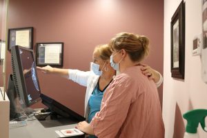
Welcome to the Diagnostic Imaging/ Radiology Department. We use a variety of cutting-edge technologies and tools to view the human body for purposes of routine check-ups/exams, diagnostics, treatments and so much more.
Most medical imaging requires a referral by your physician or other health care provider, but patients may call to schedule their own screening mammography. If you feel you need diagnostic imaging but do not have a referral, we can help you obtain a provider. Need a provider? Schedule an appointment with one of our excellent providers today.
Our Services
- Mammography
- Bone Density Testing
- Computed Tomography (CT/CAT Scan)
- Echocardiogram
- Magnetic Resonance Imaging (MRI)
- Ultrasound Imaging
- Vascular Imaging
- X-Ray Exams
To schedule an appointment please call (603) 542-3478. For more information about any medical imaging service and financial assistance options call (603) 543-6947.
Mammography
We use the latest 3D breast imaging technology to examine breast tissue one layer at a time. Did you know? 1 in 8 women will be diagnosed with breast cancer in their lifetime. A yearly mammogram screening is recommended to detect breast cancer in its earliest, most treatable stages. A mammogram can find breast cancer that is too small to be seen or felt. If you’re uninsured and cannot afford a mammogram, please call (603) 543-6947 for more information about financial assistance.

Bone Density Testing
A variety of factors contribute to your bone density including age, gender, genetics and lifestyle. Did you know 54 million Americans have osteoporosis and low bone mass? It is not uncommon for people to unknowingly live with low bone density until a fall or mild stressor causes a fracture. Osteoporosis occurs when the creation of new bone can’t keep up with the loss of old bone.
A dexa scan (bone density test) can diagnose osteoporosis by measuring both the spine and hip—two areas of the body at high-risk for fractures. This bone density test is safe, painless and non-confining, making it easier if you are claustrophobic. The test lasts approximately 20 minutes. Our team is here to assist in diagnosing and managing this disease.
Computed Tomography (CT/CAT Scan)
Computed Tomography (CT), also known as a CAT scan, is a sophisticated imaging technique that rotates around the patient to create detailed images of internal organs, bones, blood vessels and soft tissues. In an emergency these images can be used to quickly find a bleed or internal injury. These enhanced images provide superior detail and greatly improve a physician’s ability to diagnose many conditions including the size and location of a tumor.
Echocardiogram
An Echocardiogram uses ultrasound to determine function of your heart valves and chambers.
Magnetic Resonance Imaging (MRI)
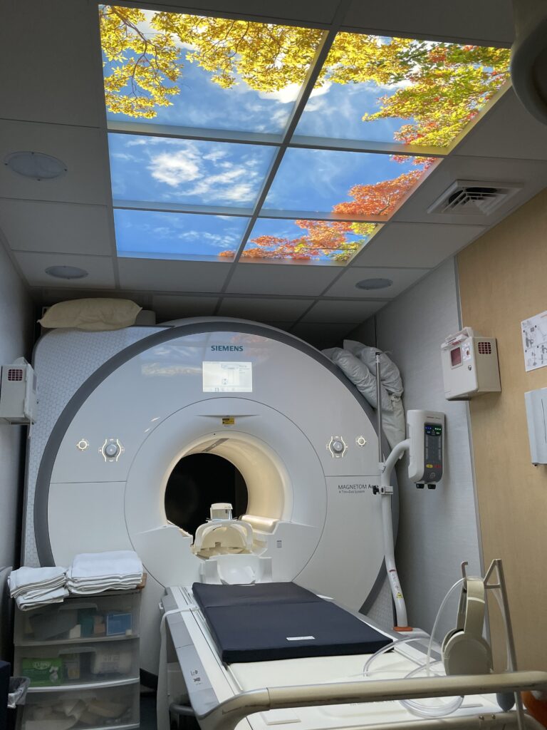
MRI exams use a magnetic field and radio waves to produce more detailed images of the body’s internal structures. Our Siemens Megnetrom Aera 1.5 Tesla with its beautifully lighted picturesque ceiling is painless, non-invasive, and free of radiation. This diagnostic tool is frequently used to detect inflammation, infection, bleeding, or abnormal masses. MRI scans are also effective in testing for neurological diseases of the brain, spine and pelvis. Due to its ability to capture details of soft tissue, MRI may also be used to view injuries to the knee, shoulder, hip, elbow, or wrist.

Ultrasound
Ultrasound is a safe, radiation-free imaging procedure that uses sound waves to visualize soft tissue and fluids in the body. It is particularly useful in examining the organs in the abdomen and pelvis (liver, gall bladder, pancreas, kidneys, spleen, bladder, uterus, ovaries and testicles). It is also used to evaluate fetal development, certain breast conditions, and the thyroid gland.
Vascular Imaging
A Vascular Imaging test examines blood flow in the major arteries and veins of the arms and legs. Ultrasound creates a series of images, allowing the size of the blood vessels to be measured. The test also measures your blood flow patterns and speed to determine if there are diseased areas in your vessels.
X-Ray Exams
X-ray or radiologic exams, use a low dose form of radiation, which passes through objects, and produces a digital image, much like a photograph of the body’s internal structure. This simple and painless procedure allows physicians to view bones, organs, or soft tissue anatomy as part of a diagnostic evaluation. X-Rays usually take 15 to 30 minutes to complete.
Please visit RadiologyInfo.org for more information about radiology and medical imaging.
